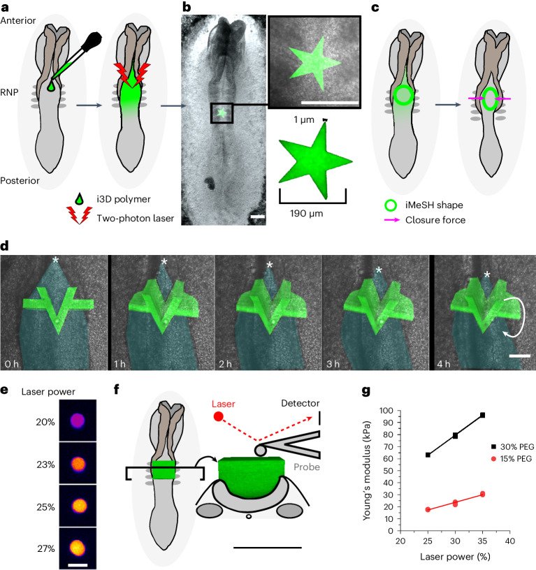
Morphogenesis requires embryonic cells to generate forces and perform mechanical work to shape their tissues. Malfunctioning of these force fields can lead to congenital malformations. Understanding these dynamic processes requires quantifying and profiling three-dimensional mechanics during vertebrate morphogenesis. Here, we describe micrometer-scale spring-like elastic force sensors, created through intravital three-dimensional bioprinting directly within the closing neural tubes of developing chicken embryos. Integrating readings from calibrated sensors with computational mechanical modeling enables the direct quantification of forces and of the mechanical work performed by embryonic tissues. As the two halves of the neural tube move toward the embryonic midline, they experience a compression exceeding one hundred nanonewtons during the apposition of the neural folds. Pharmacological inhibition of Rho-associated kinase to reduce closing force reveals the presence of active anti-closure forces that progressively widen the neural tube and must be overcome to achieve successful closure. Overall, our approach and findings highlight the intricate interplay between mechanical forces and tissue morphogenesis.