The Histology and Histopathology Facility was established to enable in-depth exploration of tissue and cellular morphology. Thanks to advanced instrumentation, our researchers can study the structure and function of cells and tissues, identify abnormalities, and contribute to the development of new therapeutic approaches.
Instrumentation
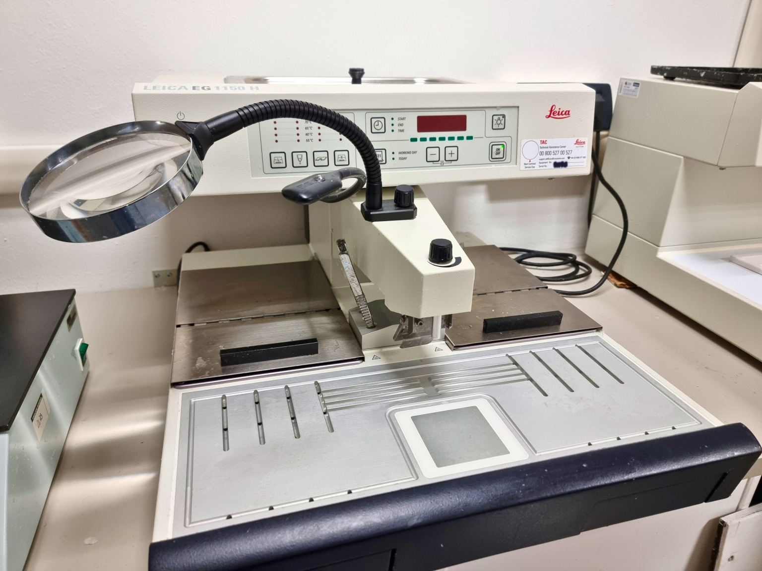
Water Baths for Slide Stretching
Instrument used for dehydration and paraffin embedding of tissue samples.
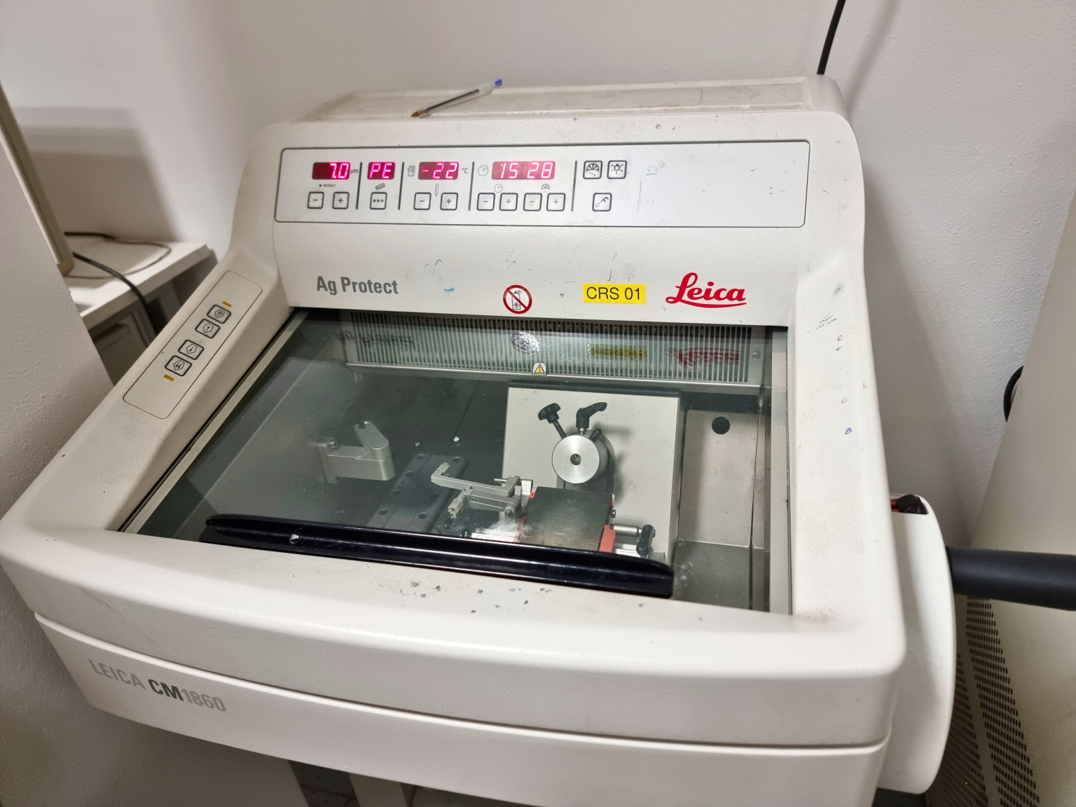
Cryostats
Leica 1860, Leica CM1860UV, and Leica CM1950 cryostats are used for cutting frozen tissue sections — essential for rapid analyses or for preserving sensitive antigens.
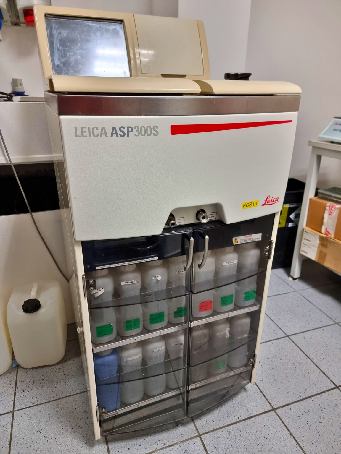
Tissue Processor
Instrument used for dehydration and paraffin embedding of tissue samples.
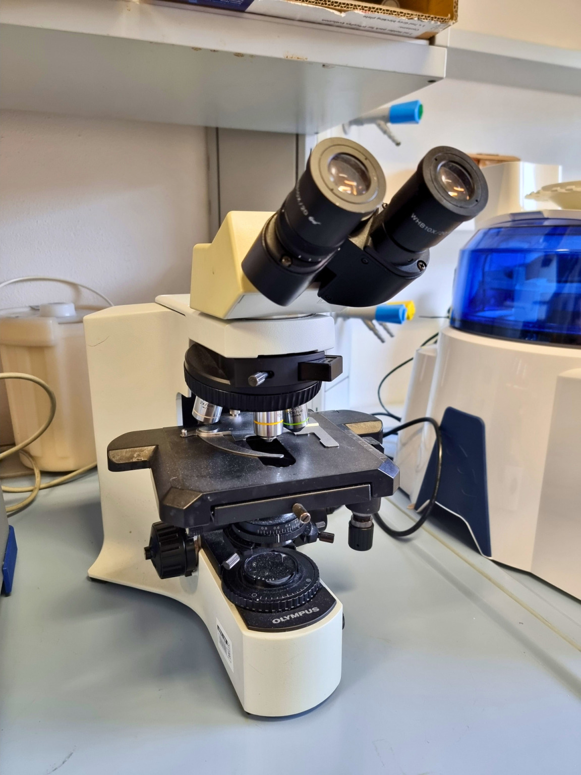
Bench Microscopes
Microscopy systems for the preliminary evaluation of tissue sections and stainings.
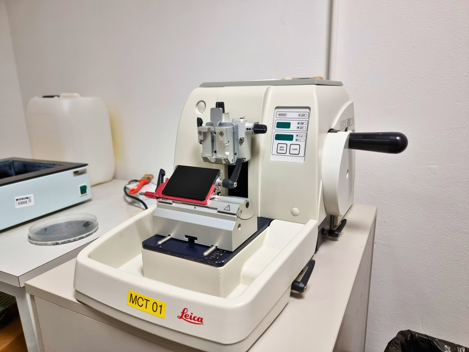
Microtomes
Instruments for cutting thin sections (3–10 μm) from paraffin-embedded tissue samples.